
Pterygium Surgery

Author: Doç. Dr. Elif Ceren Yeşilkaya, Ophthalmologist, Cornea and Refractive Surgery
Published: · Last updated:
Pterygium surgery removes tissue growing onto the cornea and covers the area with a graft of your own conjunctiva to lower the chance of regrowth.
Quick Answer
A pterygium is a benign, wedge-shaped growth that starts on the conjunctiva, the thin membrane over the white of the eye, and spreads onto the cornea. It is linked to UV exposure and is removed surgically when it affects vision or causes constant irritation.
What is a pterygium?
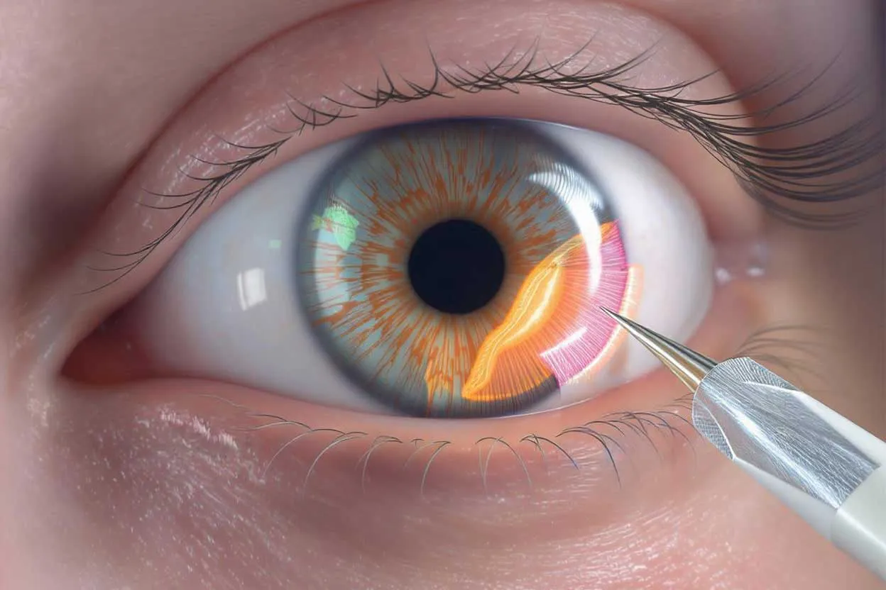
A pterygium is a fleshy, wedge-shaped growth with visible blood vessels. It starts on the conjunctiva, the thin membrane covering the white of the eye, and slowly creeps onto the cornea, the clear window at the front. It usually appears on the side nearest the nose and can affect both eyes. It is benign, but as it moves further across the cornea it can start to interfere with vision.
Ultraviolet (UV) light from the sun is the main trigger, which is why it is sometimes called surfer’s eye. Wind, dust and dry air keep the surface of the eye irritated and make things worse. People who work outdoors or live in sunny, windy climates develop it more often.
A pinguecula looks similar at first glance: a yellowish bump on the conjunctiva that stays there and does not grow onto the cornea. A look through a slit-lamp microscope is enough to tell the two apart.
When does a pterygium need surgery?
A small pterygium that has stayed the same size for years does not always need an operation. Lubricating drops and sunglasses are often enough, with a short course of anti-inflammatory drops when it flares up. Drops relieve symptoms, though they will not make the tissue smaller.
Surgery is usually considered when:
- the pterygium is moving towards the centre of the cornea, in line with your vision
- it pulls on the cornea and causes astigmatism, blurring your sight
- redness, burning, stinging or a gritty feeling persists despite drops
- it has grown noticeably in a short time
- it has come back after previous surgery
- it makes contact lenses uncomfortable or impossible to wear
- its appearance bothers you
Timing matters for the result. The further the tissue has spread across the cornea, the more visible the scar it can leave behind once removed. A pterygium approaching the pupil is therefore not left for long.
What happens at the examination?
Using the slit lamp, the doctor measures how many millimetres the pterygium has advanced onto the cornea, how thick it is and how many vessels it carries. Corneal topography maps the shape of the corneal surface. It shows whether the pterygium has caused astigmatism, meaning the cornea curves more steeply in one direction than another. Tear film tests check for dry eye, which can worsen symptoms and slow healing after surgery.
If you are also planning cataract or laser surgery, the order of treatment needs discussing. A pterygium on the cornea can distort the measurements used to calculate lens power or plan laser treatment. In that case the pterygium is usually removed first, and the measurements are repeated once the cornea has settled. Assoc. Prof. Dr. Elif Ceren Yeşilkaya decides the sequence based on what the examination shows.
How is pterygium surgery done?
The pterygium is lifted off the cornea and conjunctiva and removed, and the corneal surface is smoothed. The bare area left behind is then covered. During the operation you may feel touch or pressure.
Why a conjunctival autograft reduces regrowth
If the pterygium is cut away and the area is left bare, blood vessels grow back into the gap and the tissue often returns. With a conjunctival autograft, a thin piece of healthy conjunctiva is taken from under your upper eyelid and placed over the area where the pterygium was. Because it is your own tissue, there is no risk of rejection. The graft is held in place with fine stitches or tissue glue. The donor site heals by itself and stays hidden under the lid.
A pterygium that has already come back after surgery may need a more extensive plan, which is discussed at your examination.
Preparing for the day of surgery
Tell the doctor at your examination if you take blood thinners or any other regular medicine. Whether to pause a medicine is decided together with the doctor who prescribed it. Your eye may be covered after the operation, so do not drive, and bring someone with you.
Looking after your eye after surgery
- Redness, stinging, watering and mild aching are expected in the early days. If stitches were used, the eye may feel as though something is in it.
- Use the antibiotic and steroid drops as prescribed. Steroid drops can raise eye pressure in some people, so it is measured at your check-ups.
- The redness fades gradually as the graft settles.
While the eye heals, avoid rubbing it, wash your hands before putting in drops and keep every follow-up appointment. Your doctor sets the dates of your check-ups.
Sun protection after surgery
The conditions that caused the pterygium have not gone away, and UV exposure is one of the factors that raises the chance of regrowth. Wear sunglasses with a UV filter whenever you are outdoors. Close-fitting, wraparound frames also keep out wind and dust, and a wide-brimmed hat adds further protection. UV passes through cloud, so wear the glasses on overcast days too.
Regrowth usually shows up in the first few months as new redness and vessels at the same spot, reaching towards the cornea. If you notice this, book an appointment without waiting for your next scheduled check.
Risks and when to seek help
The most common problem is the pterygium growing back. Other possible complications include swelling, shifting or shrinking of the graft, a scar on the cornea, redness at the surgical site that lasts longer than expected and, rarely, infection.
Seek help promptly if pain gets steadily worse after surgery, your vision drops suddenly, the redness spreads or you notice thick discharge from the eye.
Assessment takes place at Medicana Zincirlikuyu (Şişli, Istanbul). A slit-lamp examination and corneal topography show how far the pterygium has advanced and whether it is causing astigmatism, and the decision on surgery and its timing is based on those findings.
{ AI · preliminary guidance }
onlineLet's talk about your cornea
AI responses do not replace a medical diagnosis.
Frequently Asked Questions
-
Can eye drops make a pterygium go away?
Drops ease redness and grittiness, but they do not shrink the tissue. A small pterygium that is not growing can be monitored, with its size measured at each check-up.
-
Is a pterygium a form of cancer?
No. A pterygium is a benign growth and does not spread to other parts of the body. If a lesion looks unusual or changes quickly, the removed tissue is sent for laboratory examination.
-
Can a pterygium come back after surgery?
Yes. Regrowth is common when the tissue is cut away and the area is left bare. Covering it with a graft of your own conjunctiva lowers that risk considerably. Regrowth usually starts within the first few months, which is why check-ups are more frequent during that period.
-
When can I go back to work?
It varies from person to person and is discussed in detail at your examination and the pre-operative consultation. If you work outdoors in sun, wind or dust, the timing is decided at your follow-up visit based on how the eye is healing.
-
Can I wear contact lenses after pterygium surgery?
Not until the graft has settled and you have finished your drops. If the pterygium made lenses uncomfortable before, lens fit is reassessed once the eye has healed.
-
I am due to have cataract surgery. Should the pterygium be removed first?
A pterygium that has reached the cornea changes its curvature and can distort the measurements used to calculate the strength of the replacement lens. In that situation the pterygium is usually removed first and the measurements are repeated once the cornea has recovered. The order is decided at your examination.
- Published
- Last updated
Appointment
Would you like more information?
A short form about Pterygium Surgery. We reply during working hours.
Treatments often evaluated together
-
Cataract Surgery
How cataract surgery works: the measurements taken beforehand, phaco step by step, lens choice, aftercare and YAG laser for later haze.
-
Dry Eye Treatment
Causes of dry eye, the tear tests done at examination, choosing artificial tears, lid hygiene, punctal plugs and dry eye checks before laser surgery.
-
Contact Lens Fitting
Soft, toric, multifocal, rigid gas permeable and scleral lenses, lens options for keratoconus, lens hygiene and the warning signs of infection.
The first step is an examination.
Your details reach Dr. Yeşilkaya's team, and we will get back to you during working hours.
+90 537 858 78 07