
Corneal Transplant

Author: Doç. Dr. Elif Ceren Yeşilkaya, Ophthalmologist, Cornea and Refractive Surgery
Published: · Last updated:
When a corneal transplant is needed: full-thickness (PK) versus layered grafts (DALK, DMEK/DSAEK), the eye bank, rejection signs and long follow-up.
Quick Answer
A corneal transplant (keratoplasty) replaces a damaged cornea, either all of it or only the diseased layer, with healthy donated corneal tissue. It is considered when other treatments cannot restore adequate vision.
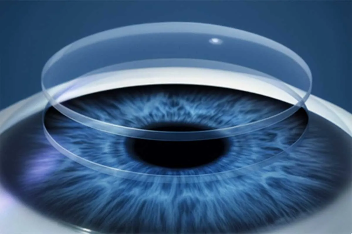
When is a corneal transplant needed?
The cornea is the clear window at the front of the eye. When it loses its clarity or its smooth dome shape, vision can deteriorate badly. A corneal transplant (keratoplasty) removes the damaged tissue and replaces it with healthy donated corneal tissue. It is considered when other treatments cannot restore adequate vision, for example in:
- Advanced keratoconus, where the cornea has become very thin or badly misshapen
- Corneal injuries that have caused serious tissue damage
- Deep corneal infections or ulcers that have left permanent damage
- Corneal dystrophies, inherited or degenerative conditions in which the cornea loses its clarity
- Corneal scars that significantly reduce vision
- Persistent corneal swelling that develops when the inner layer of the cornea fails after eye surgery
Layers of the cornea and types of transplant
The cornea is only about half a millimetre thick, but it is built in layers. On the outside is a thin covering layer (the epithelium), in the middle the stroma, which makes up most of the thickness, and on the inside the endothelium, a single layer of cells. The endothelium pumps excess fluid out of the cornea to keep it clear, and endothelial cells that are lost are not replaced. The type of transplant is chosen according to which layer is diseased.
Full-thickness transplant (penetrating keratoplasty, PK)
A round disc containing every layer is removed from the centre of the cornea, and a donor cornea of the same diameter is stitched in its place with very fine sutures. PK is needed when disease affects the full thickness of the cornea, such as deep scarring, a perforated cornea, or damage to both the stroma and the endothelium.
Because the donor’s endothelium is transplanted too, the risk of rejection is higher than with layered grafts. Vision settles more slowly than after a layered graft, stitches are removed in stages, and significant astigmatism can remain afterwards.
Front-layer transplant (DALK)
In deep anterior lamellar keratoplasty (DALK), only the epithelium and stroma are replaced. Your own endothelium, together with the thin membrane that carries it (Descemet’s membrane), stays in place. DALK is often preferred for keratoconus and for scars in the stroma when the endothelium is healthy.
Since your own endothelium is kept, endothelial rejection, the type that threatens vision most, cannot occur, and the wall of the eye remains stronger. Stitch follow-up is similar to PK. If the inner membrane tears during surgery, the surgeon may switch to a full-thickness transplant.
Back-layer transplants (DMEK, DSAEK)
When the problem lies only in the endothelium, only the inner layer is replaced. Fuchs endothelial dystrophy and persistent corneal swelling after cataract surgery fall into this group. The diseased endothelium is peeled away, and the donor tissue is passed into the eye through a small incision and pressed against the back of the cornea with a bubble of air or gas.
In DSAEK the graft includes a thin layer of stroma. In DMEK only Descemet’s membrane and endothelium are transplanted, so the tissue is much thinner. Few or no stitches are needed, and vision recovers much faster than after PK. You will be asked to lie on your back for a set time after surgery so the bubble can hold the graft in position. If the graft comes loose, more air may need to be injected into the eye.
Where does the donor cornea come from?
Donor corneas come from people who agreed to tissue donation after death and are stored at an eye bank. Before use, the donor’s blood is screened for infectious diseases such as HIV, hepatitis B and C, and syphilis, and the cornea’s clarity and endothelial cell count are assessed under a microscope.
The cornea has no blood vessels, so routine tissue matching of the kind used in kidney transplants is generally not required. The date of surgery is set according to when suitable tissue is available from the bank.
Looking after your eye after the operation
In the early days the eye may be painful, gritty, red and blurred. How long recovery takes depends on the type of transplant and varies from person to person; your doctor goes through it with you before surgery.
While the eye heals:
- Use your antibiotic and steroid (cortisone) drops exactly as scheduled.
- Do not rub the eye, and wear a protective shield at night.
- Keep water and soap out of the eye, and avoid dusty places and swimming pools.
- Avoid activities where the eye could be knocked.
- Attend every check-up, as infection and rejection are monitored at these visits.
Signs of graft rejection
The immune system can recognise donor tissue as foreign and attack it; this is graft rejection. It is most common in the first years but can happen many years after surgery. Eyes with blood vessels growing into the cornea, or that have rejected a graft before, are at higher risk.
If any of the following start, seek help the same day instead of waiting for your next appointment:
- Redness: New or increasing redness of the eye
- Light sensitivity: Discomfort in light, difficulty keeping the eye open
- Reduced vision: Blurring or misting
- Pain: Soreness or a deep ache
Rejection caught early can often be reversed with intensive steroid drops. If treatment is delayed, the graft can lose its clarity permanently, and a new transplant may be needed. Discharge and sudden loss of vision also need to be seen without delay.
Long-term follow-up: why check-ups continue for years
- Drops: Steroid drops are reduced gradually on a schedule your doctor sets, and some people stay on a low dose long term. Steroids can raise eye pressure and cause cataract, so your eye pressure is measured at every visit.
- Stitches: With a sutured graft, stitches are removed in stages at your check-ups, in an order designed to reduce astigmatism. A loose or broken stitch raises the risk of infection and rejection. If you feel grittiness or something in the eye, do not wait for your next appointment.
- Wall of the eye: After a full-thickness transplant, the graft edge stays weaker than normal cornea for life. Wear protective eyewear for sports with a risk of impact.
- Vision: Once vision has settled you may need glasses or contact lenses. Irregular astigmatism after a transplant is often managed with rigid or scleral lenses (contact lens fitting).
Which type of transplant suits your eye is decided using corneal topography, corneal thickness measurement and an endothelial cell count. The assessment is carried out by Assoc. Prof. Dr. Elif Ceren Yeşilkaya at Medicana Zincirlikuyu (Şişli, Istanbul). If you have had eye surgery before or have been followed up elsewhere, bring your previous reports and a list of the drops you use to the appointment.
{ AI · preliminary guidance }
onlineLet's talk about your cornea
AI responses do not replace a medical diagnosis.
Frequently Asked Questions
-
Can my body reject the new cornea?
Yes, the immune system can react against donor tissue. Rejection is most common in the first years but can happen many years later. If redness, light sensitivity, reduced vision or pain begins, seek help the same day; rejection caught early can often be reversed with intensive drops.
-
Does the donor cornea need to be tissue-matched?
Usually not. The cornea has no blood vessels, so routine tissue matching of the kind used in kidney transplants is generally not required. Tissue from the eye bank is screened for infectious diseases, and its clarity and endothelial cell count are checked before use.
-
How soon will my vision improve?
That depends on the type of transplant. Vision usually recovers faster after DMEK and DSAEK, where only the inner layer is replaced, than after a full-thickness transplant or DALK. What to expect in your case varies from person to person and is discussed in detail at your examination and the pre-operative consultation.
-
When are the stitches removed?
With a sutured graft, stitches are removed gradually during routine check-ups, and your doctor decides when. The order is planned to reduce astigmatism, and some stitches may be left in place for longer.
-
Will I need glasses or contact lenses afterwards?
Often, yes. Once vision has settled, any remaining prescription is corrected with glasses. If irregular astigmatism remains, a rigid or scleral contact lens may give sharper vision.
-
Does a corneal transplant last for life?
Many grafts stay clear for years. Over time the donor endothelial cells can decline or rejection can occur. If a graft loses its clarity, the transplant can be repeated.
-
What should I avoid after the operation?
Do not skip your drops or rub the eye. For as long as your doctor advises, keep water and soap out of it and avoid dust and swimming pools. An eye that has had a full-thickness transplant remains more vulnerable to injury for life, so wear protective eyewear for sports with a risk of impact.
-
Which type of transplant is used for keratoconus?
In keratoconus the inner layer of the cornea is usually healthy, so DALK, which replaces only the front layers, is often preferred. A full-thickness transplant may be needed if there is deep scarring. The choice is made at the examination.
- Published
- Last updated
Appointment
Would you like more information?
A short form about Corneal Transplant. We reply during working hours.
Treatments often evaluated together
-
Keratoconus Treatment
Keratoconus usually starts in the teenage years. Topography follow-up, corneal cross-linking (CXL), rigid and scleral lenses, rings and transplant.
-
Contact Lens Fitting
Soft, toric, multifocal, rigid gas permeable and scleral lenses, lens options for keratoconus, lens hygiene and the warning signs of infection.
-
Cataract Surgery
How cataract surgery works: the measurements taken beforehand, phaco step by step, lens choice, aftercare and YAG laser for later haze.
The first step is an examination.
Your details reach Dr. Yeşilkaya's team, and we will get back to you during working hours.
+90 537 858 78 07