
Keratoconus Treatment

Author: Doç. Dr. Elif Ceren Yeşilkaya, Ophthalmologist, Cornea and Refractive Surgery
Published: · Last updated:
Keratoconus usually starts in the teenage years. Topography follow-up, corneal cross-linking (CXL), rigid and scleral lenses, rings and transplant.
Quick Answer
Keratoconus is a progressive condition, usually starting in the teenage years, in which the cornea thins and bulges forward into a cone. Treatment aims to halt progression with corneal cross-linking (CXL) and to restore vision with glasses, specialist contact lenses, corneal rings or, at an advanced stage, a corneal transplant.
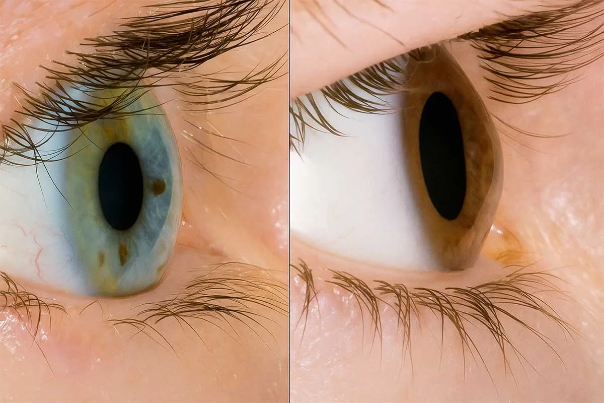
Why the cornea becomes cone-shaped
The cornea is the clear, dome-shaped window at the front of the eye and the first surface to bend incoming light. In keratoconus its tissue weakens, so the cornea thins and bulges forward into a cone. Because the surface is no longer smooth, light cannot focus properly on the retina, and images look blurred, wavy, shadowed or distorted.
Both eyes are usually affected, although one is often further along than the other. A family history of keratoconus, allergic eye disease and frequent eye rubbing all raise the risk.
Signs to watch for in teenagers
Keratoconus usually begins around puberty and can progress through the teens and twenties. The earlier it starts, the faster it tends to progress, which is why children and teenagers are followed up more closely.
- Blurred, wavy vision
- Glasses that never quite give sharp vision
- A prescription that keeps changing, particularly the astigmatism
- Light sensitivity, with halos and shadows around lights at night
- Frequent eye rubbing
A teenager whose prescription changes at every visit, or who never sees clearly with glasses, should have their cornea checked with topography.
Why stopping eye rubbing comes first
Frequent, hard rubbing puts mechanical stress on a cornea that is already weak and contributes to progression. Itching is often caused by allergy, and once the allergy is controlled with drops, the urge to rub fades too. When the itch comes, a cold compress over closed lids helps more than rubbing. This rule applies at every stage of treatment.
How keratoconus is diagnosed and monitored
Early keratoconus can go unnoticed at a routine glasses check. The key test is corneal topography (tomography), which maps the curvature of the front and back of the cornea and its thickness at every point. These maps show where the cone is, how steep it is and where the cornea is thinnest.
At follow-up visits the same scan is repeated and the maps are compared to see whether the cornea has become steeper or thinner. If it is progressing, cross-linking is considered. How often you are seen depends on your age and how quickly the disease is changing.
This is also one reason why topography is part of every assessment for laser eye surgery. Laser treatments that thin the cornea are generally not performed on a keratoconic or borderline cornea, because they can accelerate the disease.
Treatment step by step
Treatment has two goals: to stop the disease progressing and to restore useful vision. Which step suits you depends on the stage, how thick the cornea is and whether it is still changing, and many people need more than one method. Assoc. Prof. Dr. Elif Ceren Yeşilkaya plans the sequence with you at the examination, based on your scans and your age.
Glasses and contact lenses
Glasses may be enough in the early stages. As the cornea becomes more irregular, glasses stop giving clear vision and contact lenses take over. A rigid gas permeable lens creates a smooth new optical surface in front of the uneven cornea, with tears filling the gap between the two. Scleral lenses are larger; they vault over the cornea without touching it, rest on the white of the eye (the sclera) and hold a reservoir of fluid underneath. They can be more comfortable in advanced cases or for people who cannot tolerate rigid lenses.
Lenses improve vision but do not stop the disease from progressing. Fitting and trial lenses are covered on the contact lens fitting page.
Corneal cross-linking (CXL)
Cross-linking is the treatment used to halt progression. Riboflavin (vitamin B2) drops are soaked into the cornea, which is then exposed to a controlled dose of ultraviolet A light. Together, the light and riboflavin create new bonds between the collagen fibres of the cornea. The cornea becomes stiffer, and the cone stops steepening or slows down.
In the standard method the thin outer layer of the cornea (the epithelium) is removed, so the early days bring pain, grittiness, watering and blurred vision. A protective contact lens stays in place until the surface heals. The cornea stiffens gradually, and vision settles over time. Follow-up topography confirms whether progression has stopped.
CXL does not reverse the existing cone, and you may still need glasses or contact lenses afterwards. If the cornea is very thin, the standard method may not be possible; thickness measurements show this.
Corneal ring segments
At a moderate stage, if contact lenses do not give adequate vision or cannot be worn, thin, clear plastic crescent-shaped segments can be placed inside the cornea. They sit in a tunnel made in the corneal tissue, either with a laser or mechanically, and flatten the central part of the cone so the surface becomes more regular. They can be removed if necessary.
How quickly vision improves varies from person to person and is discussed at your examination. Rings do not stop progression, so if the disease is still advancing they are planned together with CXL.
Phakic intraocular lens (ICL)
If the cornea is stable but a high degree of short-sightedness or astigmatism remains, a thin lens can be placed inside the eye without removing your natural lens. The procedure is described on the phakic intraocular lens (ICL) page.
Advanced stage: corneal transplant
A corneal transplant may be needed if the cornea has become very thin, if a scar has formed in its centre, or if no contact lens gives adequate vision. In keratoconus the inner layer of the cornea (the endothelium) is usually healthy, so a transplant that replaces only the front layers, called DALK, is often preferred; a full-thickness transplant is done if needed. Recovery happens in stages, with stitches removed and vision checked at follow-up visits.
When keratoconus needs urgent attention
In advanced keratoconus the inner membrane of the cornea can tear. The cornea takes up fluid and suddenly turns white, vision drops sharply, and pain, watering and light sensitivity follow. This is called acute hydrops and needs to be seen promptly. In the days after CXL, pain that increases instead of easing, discharge or worsening vision can signal infection. In any of these situations, do not wait for your next appointment; seek help without delay.
Keratoconus follow-up and treatment take place at Medicana Zincirlikuyu (Şişli, Istanbul). At the first visit your corneal topography and corneal thickness are measured. If you wear contact lenses, they can temporarily alter the shape of the cornea and affect the scan, so ask how many days beforehand to stop wearing them when you book.
{ AI · preliminary guidance }
onlineLet's talk about your cornea
AI responses do not replace a medical diagnosis.
Frequently Asked Questions
-
Can keratoconus make me lose my sight?
Keratoconus does not affect the retina or the optic nerve. Vision loss comes from the distorted shape of the cornea and, at advanced stages, from scarring. Depending on the stage, glasses, rigid or scleral lenses, corneal rings or a transplant are used to restore vision.
-
How long does keratoconus keep progressing?
It usually starts in the teenage years and progresses through the twenties, generally slowing in the thirties and forties. The course varies from person to person, and the only way to tell whether it is progressing is to compare corneal topography scans taken over time.
-
Will cross-linking (CXL) improve my vision?
The aim of CXL is to strengthen the cornea and stop progression. In some eyes the cornea flattens slightly and vision improves a little, but the existing distortion remains. You may still need glasses or contact lenses afterwards.
-
When can I go back to school or work after CXL?
Expect pain, grittiness, watering and blurred vision in the early days, with a protective contact lens in place until the surface heals. When you can go back varies from person to person, so it is discussed at your examination, and the exact date is set at your check-up.
-
Can I have laser eye surgery if I have keratoconus?
Laser treatments that thin the cornea are generally not performed on a keratoconic eye, as they can speed up the disease. If a high prescription remains once the cornea is stable, a phakic intraocular lens (ICL) may be considered.
-
Does keratoconus run in families?
The risk is higher if a relative has keratoconus. For this reason, siblings and children of someone diagnosed with it are advised to have corneal topography as well.
-
What is the difference between rigid and scleral lenses?
Rigid gas permeable lenses sit on the cornea and create a smooth optical surface in front of the irregular one. Scleral lenses are larger; they vault over the cornea, rest on the white of the eye and hold a layer of fluid underneath. They can be more comfortable in advanced keratoconus or for people who cannot get used to rigid lenses.
-
Are corneal rings permanent?
Rings can stay in the eye permanently, but they can also be removed or exchanged if needed. Since they do not stop progression, they are combined with CXL when the disease is still advancing.
- Published
- Last updated
Appointment
Would you like more information?
A short form about Keratoconus Treatment. We reply during working hours.
Treatments often evaluated together
-
Corneal Transplant
When a corneal transplant is needed: full-thickness (PK) versus layered grafts (DALK, DMEK/DSAEK), the eye bank, rejection signs and long follow-up.
-
Contact Lens Fitting
Soft, toric, multifocal, rigid gas permeable and scleral lenses, lens options for keratoconus, lens hygiene and the warning signs of infection.
-
Phakic Intraocular Lens (ICL)
How an ICL corrects vision in eyes unsuitable for laser because of a thin cornea or high prescription, the measurements it needs and how to look after your eye afterwards.
The first step is an examination.
Your details reach Dr. Yeşilkaya's team, and we will get back to you during working hours.
+90 537 858 78 07